Every year for the last decade, the National Science Foundation has teamed up with the journal Science to co-sponsor the International Science and Engineering Visualization Challenge. The competition recognizes scientists and artists who use visual media to communicate, and promote the understanding of, scientific research.
The winners were just announced, and this year’s top visualizations are — like every year — fantastic.
Categories in this year’s competition included photography, illustration, video, posters, games and apps. Below, we’ve featured a few standout photographs and illustrations, along with this year’s first place winner from the video category — a remarkable short that combines illustration, 3D renderings, and live-action video to describe the science of the heart.
Image titles and credits listed below; all image captions via AAAS
Illustration: First Place
Title: “Connectivity of a Cognitive Computer Based on the Macaque Brain”
Credit: Emmett McQuinn, Theodore M. Wong, Pallab Datta, Myron D. Flickner, Raghavendra
Singh, Steven K. Esser, Rathinakumar Appuswamy, William P. Risk, and Dharmendra S.
Modha; IBM Research – Almaden
Cognitive Computing researchers at IBM are developing a new generation of “neuro-synaptic” computer chips inspired by the organization and function of the brain. For guidance into how to connect many such chips in a large brain-like network, they turn to a “wiring diagram” of the monkey brain as represented by the CoCoMac database. In a simulation designed to test techniques for constructing such networks, a model was created comprising 4173 neuro-synaptic “cores” representing the 77 largest regions in the macaque brain. The 320749 connections between the regions were assigned based on the CoCoMac wiring diagram. This visualization is of the resulting core-to-core connectivity graph. Each core is represented as an individual point along the ring; their arrangement into local clusters reflects their assignment to the 77 regions. Arcs are drawn from a source core to a destination core with an edge color defined by the color assigned to the source core.
Illlustration: Honorable Mention and People’s Choice
Title: “Cerebral Infiltration”
Credit: Maxime Chamberland, David Fortin, and Maxime Descoteaux; Sherbrooke Connectivity Imaging Lab
The image is the result of fiber tractography from diffusion-weighted magnetic resonance imaging. It illustrates the white matter of the brain, or in other words, its structural connections. The red smooth surface represents a glioblastoma tumor. We can see the effect of repulsion and infiltration of this mass on the white matter fiber pathways. A distance colormap is used for interpretation. Blue fibers mean that they are located within a safe distance of the tumor whereas red fibers are in a close perimeter to the tumor, and can cause severe post-operation deficits, if resected.
Photography: First Place and People’s Choice
Title: “Biomineral Single Crystals”
Credit: Pupa U.P.A. Gilbert and Christopher E. Killian; University of Wisconsin-Madison
Biomineral crystals found in a sea urchin tooth. Geologic or synthetic mineral crystals usually have flat faces and sharp edges, whereas biomineral crystals can have strikingly uncommon forms that have evolved to enhance function. The image here was captured using environmental scanning electron microscopy and false-colored. Each color highlights a continuous singlecrystal of calcite (CaCO3) made by the sea urchin Arbacia punctulata, at the forming end of one of its teeth. Together, these biomineral crystals fill space, harden the tooth, and toughen it enough to grind rock.
Photography: Honorable Mention
Title: “Self Defense”
Credit: Kai-hung Fung, Pamela Youde Nethersole Eastern Hospital (Hong Kong)
Evolution encourages diversity, allowing Nature to solve problems in more than one way. This image is a 3D CT scan of a clam and a whelk, both alive. The clam (left) is nestled comfortably in the bottom half of its shell. Note the simplicity of the hinge design in its bivalve shell. By closing the shell rapidly, the clam is able to fence off a potential attack. Yet the whelk’s shell (right) is even more amazing. The sophisticated spiral construction is astonishingly complex and strong, an architectural marvel by itself and an evolutionary success! Once the whelk slipped back into the spiral tunnel of its shell, the shell provides protection similar to a fortress. Both the clam and the whelk solve the vital problem of self defense, albeit in different ways. The whelk however has the upper hand because it has the ability to drill a hole directly through the clam’s shell by softening it with secretions and then consumes the clam as meal.
Photography: Honorable Mention
Title: “X-ray micro-radiography and microscopy of seeds”
Credit: Viktor Sykora; First Faculty of Medicine, Charles University, and Institute of
Experimental and Applied Physics, Czech Technical University; Jan Zemlicka, Frantisek Krejci,
and Jan Jakubek; Institute of Experimental and Applied Physics, Czech Technical University
High-resolution high-contrast X-ray radiography of plant seeds combined with images taken by microscopy. The X-ray images were measured using combination of a micro-focus X-ray source and a state-of-the-art hybrid pixel semiconductor detector. The detector enables imaging in socalled single photon counting regime allowing acquiring radiographs with theoretically unlimited dynamic range (in practise limited just by the number of detected photons). In combination with point-like source magnifying geometry, the technique presents a powerful tool allowing nondestructive investigation of mm-sized object of any kind. The results show a novel application of the technique to plant biology, namely the visualisation of seeds (typically 3 mm in size). For better interpretation of imaged features, the radiographs are combined with the images taken by microscopy.
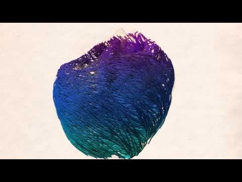
Video: First Place and People’s Choice
Title: “Alya Red: A Computational Heart”
Credit: Guillermo Marin, Fernando Cucchietti, Mariano Vazquez, Carlos Tripiana; Barcelona
Supercomputing Center