Researchers in Germany have have grown the innermost layer of human fallopian tubes in a lab. The new technique is offering fresh insights into this essential component of the female reproductive system, while also hinting at potential new directions for the treatment of various reproductive disorders.
The fallopian tubes, or oviducts, play a crucial role in human reproduction, but they’re susceptible to infections and certain forms of cancer. These small ducts, which measure about 4 to 6 inches (10 to 15 cm) in length, connect a woman’s ovaries to her uterus and facilitates transport of the fertilized egg. Typically, each woman has a pair of fallopian tubes, one located on each side of the uterus.
All too often, however, these tubes get infected by bacteria, which can lead to blockage and severe cases of infertility. Some transformed cells from the fallopian tubes can spread to the ovaries, giving rise to ovarian carcinoma—a dangerous form of gynecological cancer. And because it’s not easy for doctors to examine the interiors of their patients’ fallopian tubes, early detection is difficult. Frustratingly, it’s difficult to produce these biological conditions in the lab for research purposes.
“Here we report on the establishment of long-term, stable 3D organoid cultures from human fallopian tubes, indicative of the presence of adult stem cells.” –M. Kessler et al.
To overcome these limitations, researchers from the Max Planck Institute in Berlin developed a new technique to grow the inner mucosal layer of the fallopian tubes—a site where infections and cancer typically originate—in the lab. The results of their work now appear in Nature Communications.
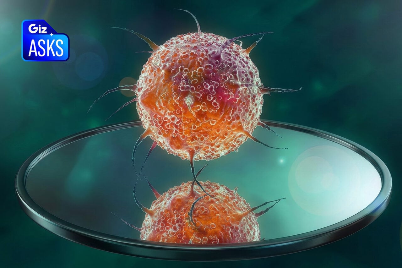
The research team, led by Thomas F. Meyer, did so by acquiring stem cells from the epithelial cells of donors, and then cultivating them under special conditions. Some of these cells transformed into “organoids,” hollow spheres consisting of many thousands of cells. These organoids were very similar to the real thing, including anatomy, structure, and biochemical processes. They even responded to hormones placed in the nutrient solution. This is good news because it likely means that these cells have the potential to grown into more specialized cells.
Remarkably, this growth occurred without any additional instruction whatsoever; it appears that the entire blueprint of fallopian tubes are stored in the epithelial cells. Also, the organoids have not experienced any “appreciable changes” in the year since they were cultivated. As the study’s lead author Mirjana Kessler explains, that is a huge advantage:
Previously available models could only keep fallopian tube epithelial cells alive for a few days. The ability to maintain the tissue-specific stem cells in culture, so they continuously replenish the cells means that these organoids can serve as research objects for much longer.
Two signalling pathways appear to be allowing for the autonomous development of the fallopian-like organoids, namely Notch and Wnt. These pathways are also known to perform key functions in embryonic development, so the researchers are keen to study this further.
These lab-grown fallopian tubes are not meant for transplant, but strictly as models for study. Looking ahead, the researchers would like to use their new ex vivo model to better understand the mechanisms of reproduction, and to learn if infections can trigger cancer.
“[Our] organoid model provides a much-needed basis for future investigations of signalling routes involved in health and disease of the fallopian tube,” conclude the researchers.
Read the entire study at Nature: “The Notch and Wnt pathways regulate stemness and differentiation in human fallopian tube organoids.”