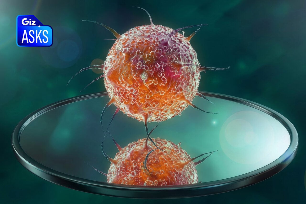
When viewed from a new angle, a different scale, or in a slightly different light, even something as familiar as the human body can look utterly alien. Case in point: these stunning anatomical illustrations, created by biomedical artist Alexandra Baker, which showcase the various nooks and crannies of the human form in all their otherworldly glory.
Illustrations like these represent science art at its finest, in that they’re not only stunning but instructive — and to anyone unfamiliar with human anatomy, they bring the added bonus of looking utterly, beautifully strange (just ask any first year med student enrolled in gross anatomy lab). Part of this obviously has to do with the fact that human physiology is just plain fascinating, but Baker’s depictions, in particular, really stuck out to us. We’ve included some examples of her work below, but you’ll find much more at COROFLOT, and at her biomedial visualization company, DNA Illustrations.
Doppler Measurement of Femoral Artery
Fibrin Clotting Factors and Inflammation
Renal Robotic Surgery
Hear Failure with Reduced Left Ventricular Ejection Fraction
Pharmaceutical Control of Parkinson’s Disease
Vegetative Growth on Mitral Valve
[DNA Illustrations via NOTCOT]