Japanese optics company Nikon has stated it is “carefully re-reviewing” information submitted by National University of Singapore optical engineer Ning Xu after commenters accused him of using AI in his winning entry to the 16th annual Small World in Motion contest.
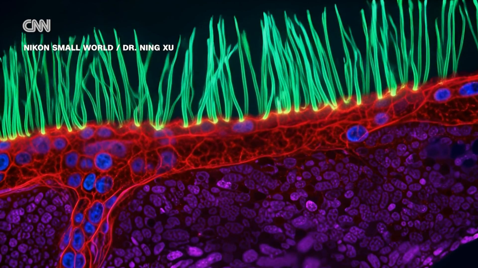
Small World in Motion bills itself as celebrating the beauty of light microscopy. According to Nature, Xu’s video submission showed the movement of cilia in lung tissue samples from a child with a chronic condition called primary ciliary dyskinesia. Unidentified red, blue, and purple structures can be seen under the cilia.
Nikon Instruments, the division of Nikon that sponsors the contest, announced Xu’s winning entry in mid-September and reposted it to LinkedIn last week. It quickly faced a storm of criticism from users who pointed out significant discrepancies. For example, University of Florida in Gainesville bioengineering researcher Edward Phelps posted to LinkedIn, “The purple structures resemble mitochondria but such a sub-epithelial structure composed of extracellular mitochondria of the same size as cell nuclei does not occur in biology.” Phelps added the blue nodules resembled nuclei but didn’t act like nuclei, and that he has no idea what the structures shown in red are.
Another LinkedIn user, UT Southwestern Medical Center PhD student Ian Donovan, pointed out that the video had a SynthID watermark, according to CNN. Those watermarks were pioneered by Google’s DeepMind research lab and have since been adopted by a large number of AI firms. Most AI detectors that attempt to spot AI via other means fare poorly, and without SynthID or equivalent watermarks, it can be difficult or impossible to determine an image’s provenance with complete certainty.
Fifth-place winner Patrick Hickey told the BBC that microscopists don’t get many chances to show off their work, which often has “very abstract and artistic qualities.”
One widely cited set of scientific guidelines allows for images of stained samples or colorized images, as long as the changes are disclosed and described. That paper was from 2010, before generative AI came along and started fabricated pixels outright.
“There’s a specific set of rules in the contest, and one of them was quite clearly that you couldn’t use generative AI to produce things and the other was obviously that it has to be taken under a microscope,” Hickey added.
In updates on LinkedIn this past weekend, Nikon said Xu was cooperating.
“Xu is respectfully complying and has provided detailed technical documentation outlining the microscopy equipment, imaging methods, and processing techniques used to create the source video,” Nikon wrote. It had previously updated a blog post on the winning entry to clarify that “An unsupervised neural-network method was subsequently used as part of the post-processing and visualization workflow to distinguish and visualize features in the grayscale data, creating a more vivid and visually engaging video.”
Xu has said as much in other LinkedIn posts, Nature noted, and said his team had used post-processing on the structures below the cilia “without making anatomical claims about what those rendered features represent.” Xu also stated that the original video of the cilia and its motion are real, and that AI was only used on the “reconstructed grayscale data to distinguish and color structures with similar morphology.”
As of Friday evening, a spokesperson for the contest organizers didn’t have any update on the situation for Gizmodo.

University of Queensland in Brisbane, Australia developmental biologist Melanie White told Nature that scientific images are data, and scientists “need to be able to trust that what we are seeing is grounded in the underlying measurement.” Former Small World in Motion judge Andrew Moore told The Telegraph that Xu’s entry made him think of “old photographs restored with AI.”
“The high-res, colorised photo may look nice, but if it was your grandma who was face-swapped, it’s going to be unsettling,” Moore told the paper.