It looks like we’re one step closer to having laser vision (or at least, laser-based diagnostic and therapeutic techniques). Researchers at Harvard Medical School and Massachusetts General Hospital in Boston have genetically engineered the world’s first “living laser.” That’s right – a living cell can shoot laser light.
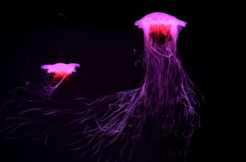
Back in 2008, scientists Martin Chalfie, Roger Tsien, and Osamu Shimomura received the Nobel Prize in chemistry for their roles in bringing Green Fluorescent Protein (also known as GFP, the protein responsible for luminosity in the jellyfish A. victoria) and its diverse applications to the front lines of scientific research.
At left you can see the laser cell. In the time between 1962 (when the protein was first isolated from A. victoria), and today, GFP has become an indispensable tool in the laboratory, with its applications ranging from that of a reporter protein to environmental biosensor to imaging tracer.
Now, GFP has been incorporated into living human cells for an entirely new purpose: the production of laser light. Optical physicists in Boston have genetically engineered a cell capable of amplifying light and emitting a bright-green directional laser beam visible to the naked eye. Their research is published in the June 12th issue of Nature Photonics.
“This is the first time that we have used biological materials to build a laser and generate light from something that is living,” said Dr. Seok-Hyun Yun, who, together with his colleague Malte Gather, created the living laser.
The principle components of a laser are:
i) A gain medium (a material with properties that allow it to amplify light)
ii) A highly reflective optical cavity (a minimum of two mirrors, between which the gain medium is placed)
iii) A means to supply energy to the gain medium (typically in the form of light).
Light passing through the gain medium is amplified, and the mirrors of the optical cavity reflect the light back through the gain medium, causing the light to be amplified repeatedly.
The above figure is from the research article, and illustrates how these three components work together to create the laser beam. The GFP fulfills the role of gain medium for the cellular laser which, when supplied with energy in the form of a weak blue light, amplifies the light in the form of an intense green laser beam. And while the width and strength of the beam may be small in comparison to traditional lasers (all of which have relied on artificial or engineered optical gain materials), it is nevertheless an order of magnitude brighter than the fluorescence observed in the jellyfish that express GFP naturally. What’s more, the genetically engineered cells remained alive, even after prolonged lasing action.
The relatively low intensity of the resulting laser beam means we still have a ways to go before we’re shooting high-power beams out of our eyes in the form of an optic blast, but Yun and Gather believe that this technology has other, less-destructive, applications in its future, including more precise imaging techniques and the targeted destruction of diseased cells:
“For light-based therapeutics, diagnosis and imaging, people think about how to deliver emission from an external laser source deep into tissue. Now we can approach this problem in another way: by amplifying light in the tissue (itself).”
Via Nature News
Research and figures via Nature Photonics