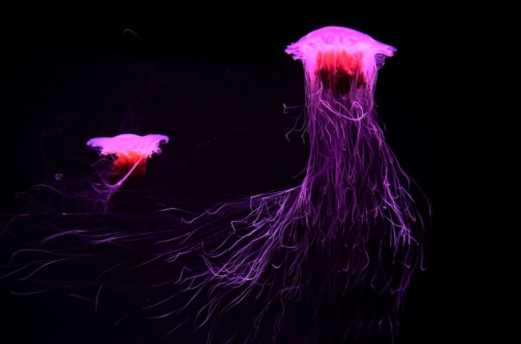
If you’ve ever been stung by a jellyfish, you’ll know how incredibly painful it is—but you might not know why. In fact, their tentacles are covered in explosive cells that are like miniature hypodermic syringes filled with venom—and in this video, you can see how they work in microscopic slow motion.
The cells, called nematocysts, fire a structure loaded with toxins outwards—a little like firing a poison dart or jabbing a hypodermic. Other cells on the jellyfish tentacle sense prey by the motion of their swimming or presence of chemicals, causing a cellular cascade which forces the nematocysts to suck in water—so much, in fact, that their venomous weapon is discharged.
Anyway, enough theory: you can see how that looks in this video put together by Destin at Smarter Everyday. With the help of a high-speed camera, a microscope and some world-class researchers from James Cook University in Australia, Destin shows us how jellyfish stings work in perhaps the best detail we’ve ever seen. So next time you get stung you may, for a few seconds at least, marvel at the wonder of nature. [Smarter Every Day]