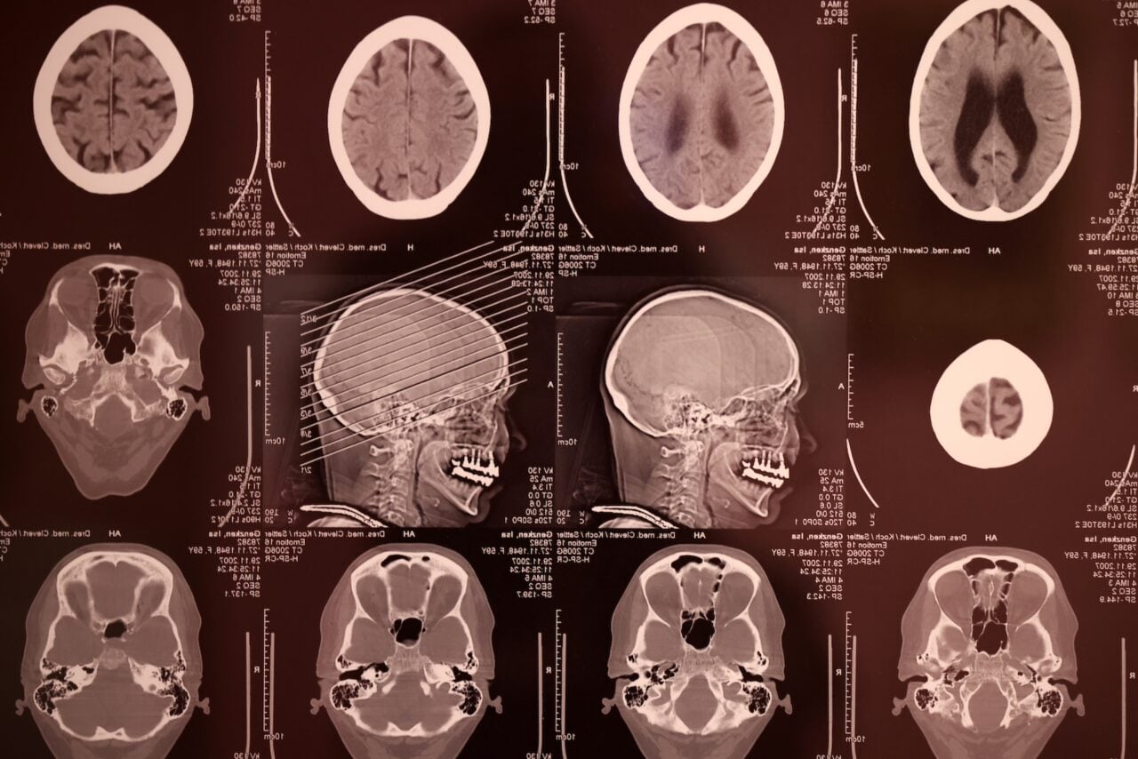
Using brain scans, neuroscientists from Emory University have revised a decades-old map of the homunculus — a visual representation of the primary motor cortex and how it corresponds to bodily awareness and control.
Top image: Black indicates neck area of motor cortex identified by Penfield in 1950. Red indicates the neck area identified by the new study.
Back in the 1940s and 50s, Canadian neurosurgeon Wilder Penfield developed the iconic motor homunculus, a distorted representation of the human body within the brain. Penfield and his team created the map by stimulating the brain with electricity in patients undergoing epilepsy surgery. From left to right, his resulting visualization showed toes and feet extending to the body’s trunk, then to a very large hand equipped with a particularly prominent thumb, followed by the head, face, and a dangling tongue beneath it all.
The new paper, published in the Journal of Neuroscience, shows that Penfield had it mostly right, save for the positioning of neck muscle control. Using functional magnetic resonance imaging, a team led by Buz Jinnah from Emory University’s School of Medicine has shown that the neck’s motor control region in the brain is actually located between the shoulders and trunk, as opposed to Penfield’s positioning in a region between areas that control the fingers and face. The new positioning more closely aligns the arrangement of the body itself.
“We can’t be that hard on Penfield, because the number of cases where he was able to study head movement was quite limited, and studying head motion as he did, by applying an electrode directly to the brain, creates some challenges,” noted Jinnah in a statement.
The team collected the fMRI data while volunteers were asked to perform various isometric muscle contractions. To prevent the participants from being able to move their heads (which would ruin the brain scans), their heads were restricted by foam padding and restraining straps.
In addition to creating a more accurate homunculus map, these findings could assist in the study of movement disorders affecting the head and neck, including head tremors and cervical dystonia.
Read the entire study at The Journal of Neuroscience: “Neural Substrates for Head Movements in Humans: A Functional Magnetic Resonance Imaging Study”.