Upon seeing this video, anaesthesiologist Professor Brian Pollard from Manchester Royal Infirmary told BBC reporter Jennifer Carpenter: “Our jaws just hit the ground. I can’t tell you the words we used as it wouldn’t be polite over the phone.”
Pollard had just seen the first-ever images of what a human brain looks like as it moves from consciousness into sleep.
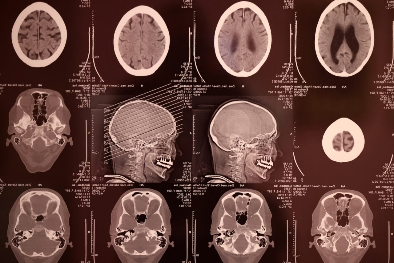
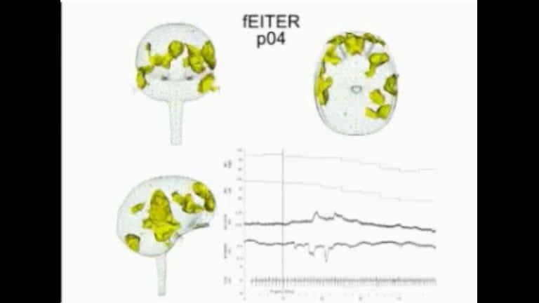
Though we’ve seen images of sleeping brains, and mapped electrical signals in the brains of people falling asleep and waking up, this is the first time scientists have used a new imaging technique called Functional Electrical Impedance Tomography by Evoke Response (fEITER). The images you see above are from fEITER, and the researchers got them by attaching electrodes to people’s heads and sending electrical currents through their skulls. The technique is good for mapping where electrical signals are zooming (or not zooming) through your brain — electrical signals in the brain interrupt currents from the electrodes, and the resulting images show us exactly which parts of the brain are active.
So, what’s the big shock? Carpenter explains:
As the patient goes under, different parts of the brain seem to be “talking” to each other, a team told the European Anaesthesiology Congress in Amsterdam . . . The finding supports a theory put forward by Professor Susan Greenfield, from the University of Oxford, that unconsciousness is a process by which different areas of the brain inhibit each other as the brain shuts down.
Obviously, a lot more research and imaging need to be done before we can be certain what these electrical patterns mean in our brains. But the more we map the synaptic byways of our brains, the closer we’ll be to re-engineering consciousness itself.
via BBC