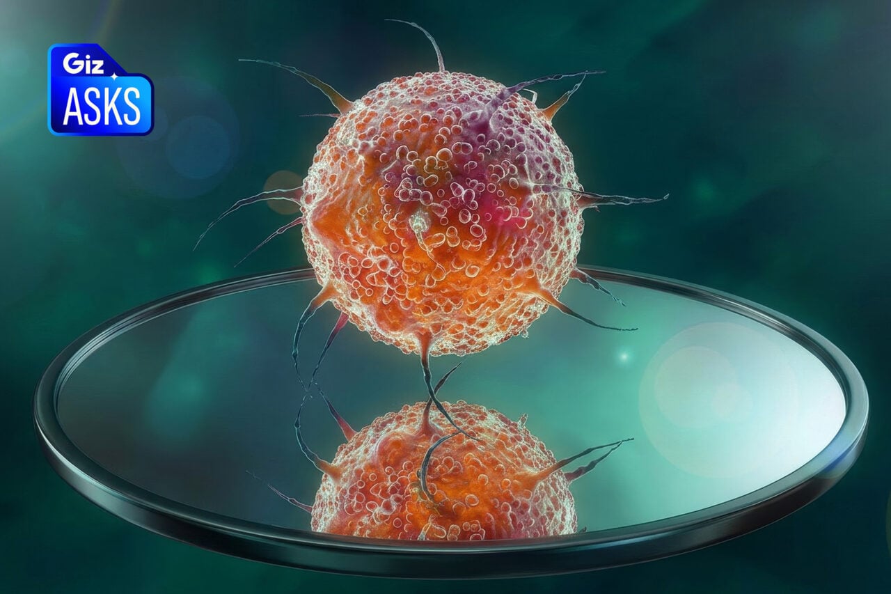
This is a stunning 3D map that shows how six feet of of DNA can be crammed inside a single chromosome — a space that’s only a hundredth of a millimeter across. Not surprisingly, it looks like something that would go well with meatballs.
Chromosomes, those packages of genetic material found in our cells, were discovered way back in the late 1800s, but scientists have struggled to understand the exact way DNA molecules fold into them across three-dimensions. But a new study conducted by researchers at MIT and the University of Massachusetts Medical school has resulted in the world’s first comprehensive model of the 3D organization of condensed human chromosomes.
https://gizmodo.com/how-artificial-chromosomes-could-transform-humanity-754993569
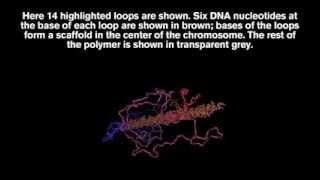
Their model shows that DNA creates tightly compressed loops that start from a flexible scaffold. The researchers, a team led by Leonid Mirny, say it’s a very efficient way of packing DNA material. During this condensed state, which can only be seen when cells are dividing, cells neatly separate and distribute their chromosomes such that each daughter cell receives the full compliment of genetic material. At other times the chromosomes are more loosely organized inside the cell nucleus.
The scientists discovered that, as cells begin to divide, chromosomes are completely reorganized. “Unlike proteins, which fold into very defined structures, the chromosomes form a completely different condensed object every time,” noted grad student Geoffrey Fudenberg in an MIT statement. “It appears similar macroscopically but the individual regions of the genome can be folded in very different ways in different cells.”
Long strands of DNA, which can reach upwards of 6.5 feet (2 m), wind around proteins called histones, giving rise to it’s “beads on a string” structure. Scientists have proposed many models to explain how those strands of millions of beads can be packed inside a chromosome, but there’s a complete lack of consensus on the matter.
The new study resolved the problem by using a technology called Hi-C, which shows the frequency of interaction for each pair of regions in the entire genome.
The Hi-C technique “provides a modern day molecular microscope, with the power to see inside of these bodies and elucidate their principles of organization,” said Nancy Kleckner, a professor of molecular and cellular biology at Harvard University — a result she described as “a new, yet satisfyingly familiar, view.”
Read the entire study at Science: “Organization of the Mitotic Chromosome.”
Top image: Mirny et al/MIT.