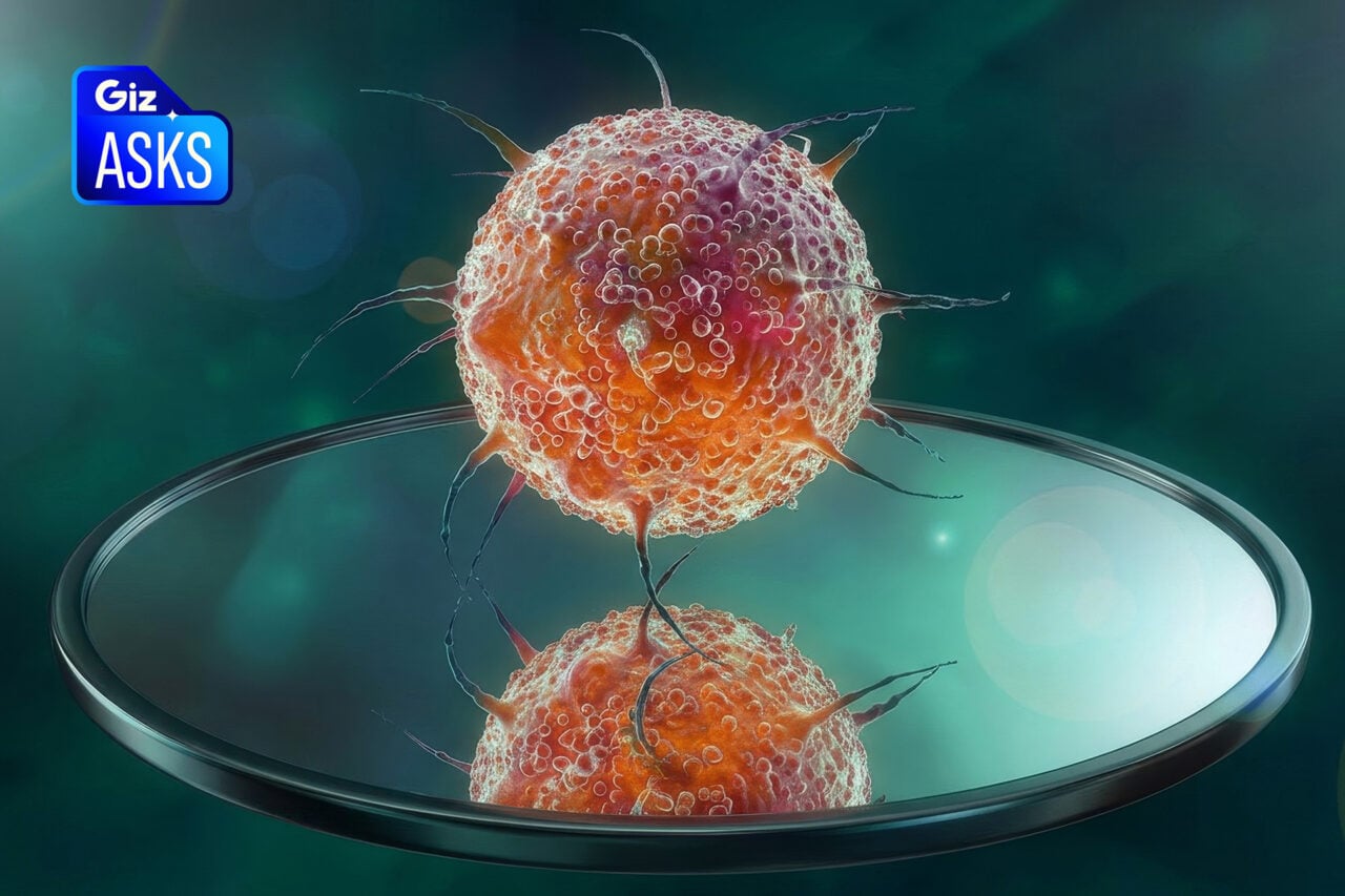
For the last 60 years, scientists have known that DNA’s structure is composed of a spiraling corkscrew. They know this thanks to molecular theory and and an old-time technique called X-ray crystallography, where patterns of dots are converted into an overarching image using mathematics. But now, using an electron microscope, scientists have taken a highly vivid snapshot of a tightly packed bundle of DNA — perhaps the best glimpse yet of this life-giving molecule.
Addendum: An earlier version of this article incorrectly stated that a single strand of DNA had been imaged — including its characteristic double-helix. Rather, the image is of a bundle of DNA molecules, and not an isolated one. In addition, the technique will not allow the researchers to study the interactions of RNA and proteins. We regret the errors.
The image was taken by Enzo di Fabrizio from the University of Genoa, Italy. He choreographed the scene by pulling small strands of DNA from a diluted solution and then propping them up like a clothesline between two nanoscopic silicon pillars.
The trick to the technique was in acquiring discrete strands of DNA that could be stretched out and ready to view with an electron microscope. Di Fabrizio managed this by creating a pattern of pillars that repelled water — which resulted in quick moisture evaporation and residual strands of DNA all ready to go.
Then, in order to create a high-resolution image, di Fabrizio drilled tiny holes in the base of the nanopillar bed and shone beams of electrons.
The result is this spectacular image — not of a single molecule of DNA — but a tightly packed bundle of them. The researchers modelled the structure as a collection of seven parallel DNA double-helices, what created a structure with the same thickness as the imaged fibre.
The paper, “Direct Imaging of DNA Fibers: The Visage of Double Helix,” was published in Nano Letters.
Supplementary source: New Scientist and The Guardian.
Images: Enzo di Fabrizio.